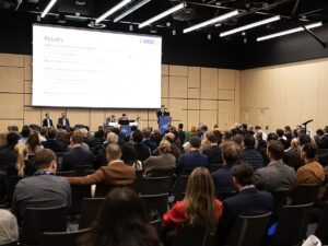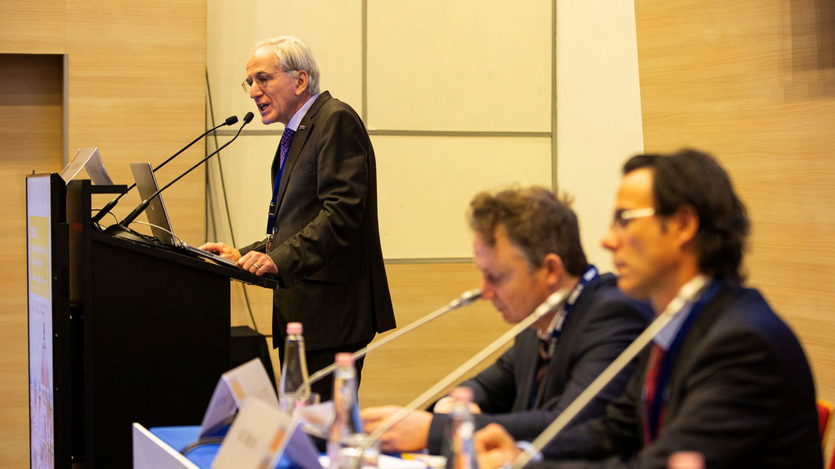
The 10th Meeting of the EAU Section of Urological Imaging got off to a good start on Thursday, 10 November, with some of the biggest names in imaging looking at recent developments and also venturing a guess as to what developments would be concrete enough to change the way urologists and radiologists work in the coming years.
ESUI22 is being held in conjunction with EMUC22, the 14th European Multidisciplinary Congress on Urological Cancers, and a variety of satellite meetings. Together, this four-day event gives an extensive update on urological cancers in a multidisciplinary perspective, featuring speakers representing ESMO, ESTRO and the EAU.
Potential of AI
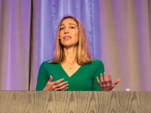
In the first plenary session “Standard today, but what about tomorrow?” radiologist Prof. Jelle Barentsz (Nijmegen, NL) presented the need for new protocols of quality control in imaging, and re-assessed the role of artificial intelligence (AI) now that its use has widened.
AI had made improvements, even in the past two years, but it still cannot compete with an expert gaze. It is however approaching the level of skill of a typical radiologist and this opens up new possibilities. According to Prof. Barentsz, AI is the radiologist’s friend and can help diagnosis in several ways. “Use of AI can shorten reporting time, and helps the radiologist with easy, more obvious cases. It can make the report and annotate the region, speeding up evaluation time. It also improves detection by offering a ‘double read’: an extra check by a computer that’s never tired.”
Importantly, the radiologist “remains in control” but AI can ease the workload, especially in the post-COVID period where medical experts are prone to burn-outs. The radiologist can focus on consulting, and has more time for creative thought. Interestingly, use of AI also allows integration with non-imaging tools and other “-omics”.
Prof. Barentsz went on to give a quick overview of his experiences with the Transurethral Ultrasound Ablation, or TULSA, procedure at the Busch Center (in Alpharetta GA, USA). Initial impressions left him “flabbergasted”, and he concluded that it will certainly play a role in the coming years as a viable treatment option.
Other developments on the horizon
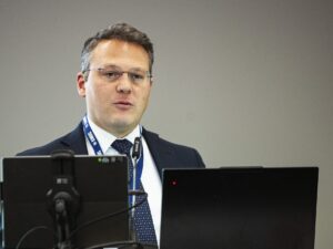
During the same session, Dr. Vincenzo Scattoni (Milan, IT) offered a urologist’s perspective on the development of TRUS over the past few decades. Dr. Scattoni concluded: “TRUS’s utility in clinical practice has been continuously confirmed over the years, but unfortunately there are no current developments that have proven to significantly improve cancer detection. Based on well-designed controlled studies, the combination of targeted biopsy schemes and systematic biopsies provides the highest detection rate.”
Prof. Frederik Giesel (Dusseldorf, DE) had a pre-recorded presentation on the different tracers that were beginning to see use in PSMA PET-CT. A particular highlight was the emergence of PSMA-ligand therapy, currently in Phase II trials and carefully starting to be included in medical guidelines: “Exciting times are waiting for us in the future of PSMA,” said Prof. Giesel.