There are several promising imaging techniques on the horizon, with the potential of changing the way oncology experts treat their patients. Novel modalities in ultrasound, combined with machine learning, further evolution of PET-CT beyond PSMA, the use of nanoparticles as a contrast agent and the emergence of the VI-RADS score are all anticipated in the near future.
EMUC congresses are typically organised to coincide with the Annual Meeting of the EAU Section for Urological Imaging (ESUI), as well as other imaging-specific workshops and meetings. With the move to an online congress, the wide imaging scientific programme was streamlined somewhat and integrated into the EMUC20 Virtual Congress.
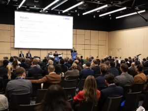
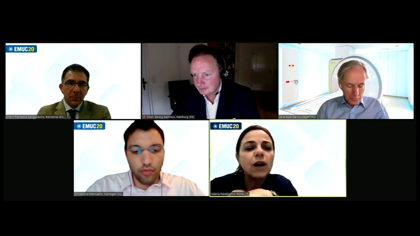
It was in the tenth plenary session, ‘Fragments of Imaging’ that most of the latest developments in oncological urology-related imaging were presented. The Saturday afternoon session was chaired by ESUI Chairman Prof. Salomon (Hamburg, DE) and Prof. Sanguedolce (Barcelona, ES) and at its peak attracted some 470 simultaneous viewers (or 630 unique viewers spread out over the entire session). Many questions for the speakers were submitted through the Q&A system, several of which were covered in the discussion following the presentations.
Ultrasound has scope for improvement
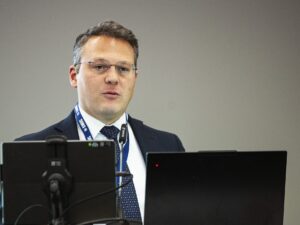
Mr. Christophe Mannaerts (Nijmegen, NL) presented the latest developments in ultrasound, specifically when compared to the mpMRI pathway. The latter poses some challenges, including “costs and accessibility, inter-reader reproducibility, inter-operator reproducibility with the necessity of cross-modality registration, and the variability in results from different centres of expertise,” said Dr. Mannaerts, citing several recent publications.
Possible benefits of the use of (improved) ultrasound in urology include reduced costs, better accessibility and easier lesion targeting. This makes ultrasound a field that is worth further developing and refining, according to Mr. Mannaerts. Improvements can be found in the use of micro-ultrasound, working at 29MHz instead of the more traditional 8-12MHz. Mannaerts: “The use of this increased frequency has led to 300% improved resolution, revealing microstructures and tissue patterns. The PRI-MUS protocol has been developed for this case.” Other potential developments include novel modalities in ultrasound, like contrast ultrasound dispersion imaging (CUDI).
Mr. Mannaerts concluded that ultrasound imaging for PCa detection and localisation is improving, and it can become a more accessible and viable modality for targeted biopsy is improvements are made in developing a standardised acquisition and reporting scheme, and high-quality benchmarking with the mpMRI pathway in the same patient group.
Other presenters in the busy session were Prof. Jurgen Futterer (Nijmegen, NL), Prof. Frederik Giesel (Heidelberg, DE), Prof. Jelle Barentsz (Nijmegen, NL), and Prof. Valeria Panebianco (Rome, IT).
New directions in imaging
Prof. Futterer summarised developments in imaging in the past thirty years, indicating that abbreviated (or “fast”) MRI prostate protocols might make for an appealing option, depending on indications, cost, experience, and the development of smart algorithms and AI. Prof. Giesel discussed the “rising star” of imaging, the new fibroblast activation protein inhibitors (FAPI-)PET/CT. Advantages compared to FDG include no need for diet or fasting for patients and quicker image acquisition after tracer application. “FAPI opens a new application for PSMA-negative PCa patients,” Prof. Giesel concluded.
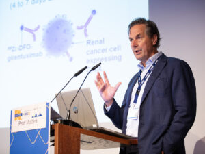
Prof. Barentsz, introduced by the moderators as “the father of the PI-RADS” presented some first results on the use of nanoparticles, and the perspective of their use as a contrast agent. Nano-MRI guided RT shows “promising results” in detecting small positive lymph nodes that PLND and PSMA PET/CT have trouble finding. Prof. Barentsz expects approval for the agent, MR-Linac, in the next two years.
“Can VI-RADS avoid TUR-B prior to radical cystectomy?” asked Prof. Panebianco in the final talk of the session. “Yes we can,” she concluded. Her presentation on the Vesical Imaging-Reporting and Data System proposed an MRI pathway for bladder cancer patient stratification: NMIBC vs MIBC, and MIBC-T2 vs Locally Advanced BC -T3–T4, only then sampling TUR for confirmatory pathology in VI-RADS 5.
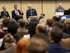
The session closed with an unfortunately brief panel discussion on delegate’s questions. Considering the enthusiasm for the session, and the hotly anticipated new imaging technologies on display, urological cancer specialists can expect big changes in their imaging options in the near future.
EMUC20 delegates can re-watch the entire session (and the rest of EMUC20) through the EMUC20 Resource Centre. Contents will be constantly updated over the coming days.