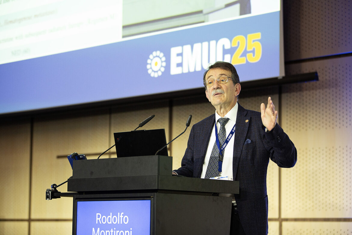
During “Plenary Session 3: Rare genitourinary tumours and unconventional pathologies” held today, pathologist Prof. Rodolfo Montironi (IT) presented a patient case and gave an overview of Wilms tumour (WT) – also known as nephroblastoma – in adult patients.
The patient was a 44-year-old female who had left-back pain, a 10 cm left kidney mass, 3.5 cm para-aortic mass, and multiple liver lesions. Fine-needle aspiration (FNA) and formalin-fixed paraffin-embedded (FFPE) cell block were performed before the operation. Presurgical therapy was applied, then the tumour was resected. The results showed it was post-therapy favourable (no anaplasia), high-risk (blastemal predominant) triphasic WT. The Children’s Oncology Group (COG) stage was IV (hematogenous metastases).
Prof. Montironi stated that WT in adult patients is a difficult and unexpected diagnosis, whereas RCC is the most difficult kidney tumour. In an adult patient, there is a much broader differential diagnosis, requiring significant additional ancillary testing to consider other possibilities, based on FNA findings and immunohistochemical and molecular studies.
Prof. Montironi enumerated the most helpful pathologic features when using FNA to diagnose WT in adult patients:
- Malignant epithelioid cells with tubular and rosette-like architecture
- Frequent mitotic figures
- Necrosis
- WT1 expression by immunohistochemistry
“The FNA in adult patients can be a bit tricky because there are lesions that can mimic [other tumours] or are difficult to diagnose. One of these is a metanephric adenoma, which is related to WT. Some authors would refer to it as the benign counterpart or the beginning of WT. We have to identify the criteria for malignancy, such as the presence of a blastemal component, mitosis, and necrosis. These indicate WT,” said Prof. Montironi.
According to him, from the morphological point of view, histology in pediatric and adult patients is identical. “The components represent stages in normal or abnormal nephrogenesis. There are undifferentiated blastemal cells, and also cells differentiating towards epithelial and stromal elements. We see three patterns, which means they are triphasic. If there are two, it is biphasic, and sometimes, it’s monophasic, which can be more difficult to diagnose.”
For more information on the patient case and WT, (re)watch the Prof. Montironi’s full presentation on the EMUC25 Resource Centre.
Medical oncologist Asst. Prof. Ronan Flippot (FR), urologist Prof. Juan Gómez Rivas (ES), pathologist Prof. Rodolfo Montironi (IT), radiotherapist Dr. Simon Spohn (DE), and radiologist Prof. Harriet Thoeny (CH) led Plenary Session 3.